Have you ever found yourself navigating through the intricacies of staining protocols while trying to achieve the perfect visualization in your microscopy studies? If so, you are certainly not alone. The world of staining techniques can be both rewarding and challenging, particularly when it comes to the use of Methylene Blue. Understanding the best practices for employing this dye can significantly enhance the clarity and accuracy of your results.

Understanding Methylene Blue
Methylene Blue is a cationic dye that has been widely used in biological staining for over a century. Its solubility in water and ability to bind to nucleic acids, proteins, and cellular structures make it a valuable tool in microscopy. The dye is particularly effective for staining cellular components and tissues, providing contrast that aids in the visualization of structures under a microscope.
Properties of Methylene Blue
Before using Methylene Blue in your staining protocols, it is essential to understand its properties. The following table summarizes key characteristics:
| Property | Description |
|---|---|
| Molecular Formula | C16H18ClN3S |
| Molecular Weight | 319.85 g/mol |
| Solubility | Soluble in water, alcohol, and glycerol |
| Color | Blue |
| pH Range | 4.0 to 6.0 |
Understanding these properties sets the foundation for developing effective staining protocols.
Importance of Methylene Blue in Staining Protocols
Methylene Blue plays a pivotal role in various staining techniques across different fields of study, including histology, microbiology, and clinical diagnostics. It enhances the visualization of cellular and subcellular structures, making it invaluable for researchers, clinicians, and educators alike.
Applications in Histology
In histology, Methylene Blue is commonly used to stain tissue sections, helping to highlight cell nuclei and other important structures. It is especially beneficial in identifying cellular morphology and understanding tissue architecture.
Role in Microbiology
When it comes to microbiology, Methylene Blue is often employed for staining bacterial cells. Its affinity for nucleic acids makes it an effective counterstain, allowing differentiation between different types of bacteria in a sample.
Preparing for Methylene Blue Staining
Before you begin a staining protocol using Methylene Blue, several preparation steps are crucial.
Selecting the Right Concentration
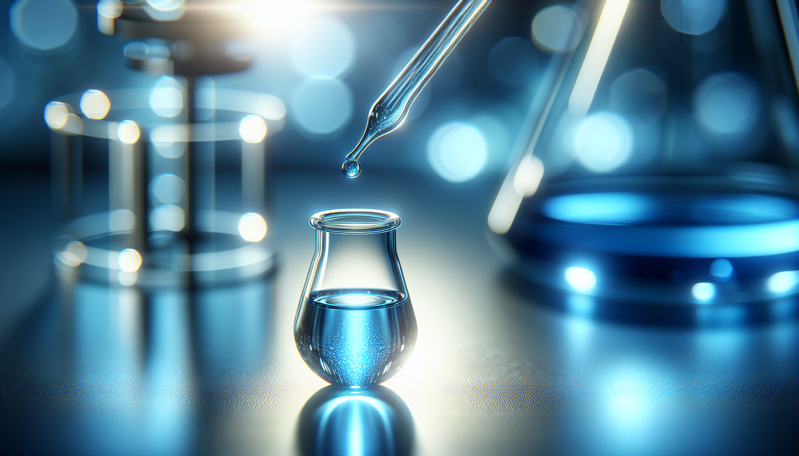
The concentration of Methylene Blue is critical to obtaining optimal staining results. Traditionally, a 0.5% to 1% aqueous solution is utilized for most applications. However, the concentration may vary depending on the specific requirements of your experiment. Testing a range of concentrations can help you determine the ideal level for your needs.
Preparing Methylene Blue Solution
-
Materials Required:
- Methylene Blue powder
- Distilled water or appropriate buffer
- pH meter or pH strips
- Glass beaker or container
-
Steps:
- Measure out the desired amount of Methylene Blue powder based on your intended volume.
- Slowly add distilled water while stirring until the powder completely dissolves.
- Adjust the pH of the solution if necessary, aiming for a pH of around 4.0 to 6.0 for optimal staining.
Safety Precautions
Safety should always be a priority when handling chemicals. Methylene Blue is generally considered safe; however, it is important to take certain precautions:
- Wear gloves and a lab coat.
- Handle the dye in a well-ventilated area.
- Be aware that Methylene Blue can stain skin and clothing, so take care when using it.
Performing the Staining Protocol
Once you have prepared your Methylene Blue solution, you can move on to the actual staining process. This section outlines a systematic approach to ensure accurate and reproducible results.
Fixation of Samples
Before staining, fixation is a critical step, particularly for tissue samples. Fixation preserves the cellular structure and prevents degradation.
-
Common Fixatives:
- Formaldehyde
- Ethanol
- Bouin’s solution
-
Procedure:
- Immerse the samples in the chosen fixative for an appropriate duration, typically between 10-60 minutes, depending on the fixative used and the sample type.
- Rinse the samples with distilled water to remove excess fixative.
Staining with Methylene Blue
- Immerse the Sample: Place your fixed sample into the Methylene Blue solution, ensuring it is fully submerged.
- Staining Time: The ideal staining time may vary; typically, 5-20 minutes is sufficient. Observing the sample periodically can help determine the optimal stain intensity.
- Rinse: After staining, rinse the sample gently with distilled water to remove any unbound dye. This step is crucial to avoid background staining.
Mounting the Samples
To visualize your stained samples under a microscope, proper mounting is essential.
-
Materials Required:
- Microscope slides
- Coverslips
- Mounting medium (if necessary)
-
Procedure:
- Place a drop of mounting medium on the slide (if using).
- Carefully position your stained sample on the slide.
- Gently place the coverslip over the sample, avoiding air bubbles.

Troubleshooting Common Issues
Despite following protocols, challenges can arise during the staining process. Here are some common issues you may encounter and how to address them.
Inconsistent Staining
If you observe inconsistent staining across samples, consider the following factors:
- Concentration of Methylene Blue: Confirm that the dye concentration is appropriate.
- Staining Time: Ensure all samples are stained for the same duration.
- Fixation Techniques: Standardize fixation protocols to maintain consistency.
Background Staining
Excessive background staining can obscure the cellular details you wish to examine. Here are some potential solutions:
- Rinse Thoroughly: Ensure unbound dye is effectively rinsed from the sample.
- Shorten Staining Time: A shorter incubation time may help reduce background staining.
Optimizing Microscopy Settings
Once staining is complete, the next step involves adjusting your microscopy settings for optimal viewing of your samples.
Selecting the Right Microscope
Different microscopy techniques yield varying results when observing stained specimens. Choose the appropriate microscope based on your requirements:
- Light Microscope: Suitable for standard observations.
- Phase Contrast Microscope: Enhances unstained and stained samples by utilizing differences in refractive index.
- Fluorescence Microscope: If using a modified Methylene Blue protocol with fluorescent tags, a fluorescence microscope is ideal.
Adjusting Illumination
Proper illumination is key to maximizing visualization:
- Brightfield: Utilize brightfield illumination for standard observations.
- Adjust Light Intensity: Modulate light intensity based on the thickness and transparency of the specimen.
Choosing the Right Objective Lens
Selecting the correct objective lens magnification is critical to your observation:
- Low Power (10x or 20x): Ideal for initial observations.
- High Power (40x or 100x): Necessary for detailed examination of cellular structures.
Preservation of Stained Samples
After successful staining and visualization, you may wish to preserve your specimens for future reference or analysis.
Long-Term Storage Conditions
- Storage Medium: Use an appropriate mounting medium that offers clarity and stability.
- Temperature: Store samples in a cool, dark place to prevent degradation.
Labeling and Documentation
For effective tracking and future analysis:
- Clearly label your samples, including the staining protocol used, date, and any pertinent observations.
- Maintain a detailed lab notebook for methodological notes, any variations made, and observations noted during the staining process.
Conclusion
Implementing best practices while using Methylene Blue in staining protocols can dramatically improve the clarity and accuracy of your microscopic observations. From the correct preparation and application techniques to careful microscopy settings and sample preservation, each step plays a significant role in ensuring optimal results.
By remaining attentive to the details of your protocols and continuously refining your techniques, you will develop a strong foundation for successful staining practices. As you engage in further studies or experiments, these guidelines can serve as a reliable reference to enhance your skills in microscopy and staining.