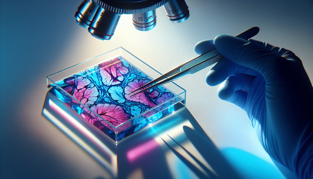
Have you ever considered the role of histological stains in understanding cellular structures and diagnosing diseases? Methylene blue is one such stain that has become invaluable in the field of histology. This guide aims to provide a comprehensive understanding of methylene blue, its applications, significance, and methodologies for effective use.

What is Methylene Blue?
Methylene Blue is a synthetic dye that has been used for over a century in various scientific applications. In histology, it serves as a vital staining agent that highlights cellular components, allowing for a clearer understanding of tissue morphology. Originally synthesized in the 19th century, its multiple applications extend beyond histology. However, its use as a stain is particularly noteworthy.
Chemical Properties of Methylene Blue
Understanding the chemical properties of methylene blue is crucial for its application in histology. It is a cationic dye, which means it carries a positive charge. This property enables it to bind effectively to negatively charged cellular components, such as nucleic acids and certain proteins. Here’s a breakdown of its chemical characteristics:
| Property | Description |
|---|---|
| Chemical formula | C16H18ClN3S |
| Molecular weight | 319.85 g/mol |
| Appearance | Dark blue crystalline powder |
| Solubility | Soluble in water, slightly soluble in alcohol |
| pH | 4.5 – 7.0 during staining |
Applications of Methylene Blue in Histology
Methylene blue’s versatility makes it suitable for various histological applications, including staining of tissues and cells, highlighting specific structures, and even aiding in physiological studies. Below are the primary applications this dye offers in histology:
1. Staining Biological Tissues
Methylene blue is primarily utilized to stain biological tissues. It is excellent for visualizing cell nucleus and cytoplasm, often resulting in a stunning contrast that makes it easier to identify different cellular components.
2. Identification of Cellular Structures
With its ability to bind to nucleic acids and certain proteins, methylene blue enhances our ability to identify and differentiate cellular structures. This is particularly useful in identifying tissue types, such as muscles, connective tissues, and epithelial cells.
3. Fast Diagnosis in Clinical Settings
In clinical settings, methylene blue has found applications in rapid diagnosis. It can be used to stain samples for immediate assessment, especially in situations where time is of the essence.
4. Observing Cellular Metabolism
Methylene blue is also noted for its role in studying cellular metabolism. In certain conditions, it can act as a redox dye, providing insight into metabolic processes in live cells.
The Mechanism of Action of Methylene Blue
Understanding how methylene blue interacts with tissues is essential for its effective use as a histological stain. The primary mechanism involves the dye’s electrostatic interactions and permeability across cellular membranes.
Binding Mechanism
The binding of methylene blue occurs through ionic interactions with negatively charged groups present in nucleic acids and proteins. When tissues are exposed to this dye, the positivity of the methylene blue allows it to penetrate the cell membrane and bind to its targets.
Staining Protocol
To effectively stain tissues or cells with methylene blue, a standardized protocol should be followed. This ensures consistent results and enhances reproducibility in your work.
-
Preparation of Slides: Begin by preparing your tissue samples on microscope slides. Ensure that they are adequately fixed and deparaffinized if necessary.
-
Dilution of Methylene Blue: Dilute methylene blue in distilled water to a concentration between 0.1% to 1%, depending on the specific application.
-
Staining Process: Immerse the slides in the prepared solution for 5-10 minutes, allowing the dye to interact with the cells.
-
Rinsing: After staining, rinse the slides with buffered saline or distilled water to remove any unbound dye.
-
Mounting: Lastly, apply a suitable mounting medium and cover slip for microscopic examination.
Advantages of Methylene Blue Staining
There are multiple advantages to utilizing methylene blue as a histological stain. Understanding these benefits can aid you in optimizing your staining protocols.
Clarity and Contrast
Methylene blue imparts a high level of clarity and contrast between different cellular structures. The unique color it produces allows for easy identification of nuclei, cytoplasmic components, and other cellular structures.
Cost-Effectiveness
One of the significant advantages of methylene blue is its cost-effectiveness. Compared to other histological stains, methylene blue is relatively inexpensive, making it a favorite among educational institutions and clinical laboratories alike.
Versatility
Methylene blue can be utilized across a range of sample types, including animal tissues, plant tissues, and even bacterial samples. Its wide applicability offers researchers and clinicians great flexibility in their work.
Safety Profile
Methylene blue is generally considered safer than many other histological stains, making it a suitable option for educational laboratories where handling precautions may be limited. While it is essential to follow standard laboratory safety protocols, methylene blue has a lower risk of causing adverse reactions.
Limitations of Methylene Blue
While methylene blue has several advantages, it also has inherent limitations that you should be aware of when employing it as a stain.
Non-Specific Staining
Methylene blue may bind to non-target cellular components, leading to non-specific staining. This can complicate the interpretation of structures and may require additional staining protocols to clarify findings.
Resolution Limits
The dye does not provide as high of a resolution as some advanced alternative stains and may fall short in detecting subtle differences in tissue morphology. Therefore, it might not be suitable for ultra-structural studies in high-resolution microscopy.
Limited Usage in Immunohistochemistry
Immunohistochemistry often relies on more specific markers and stains tailored to particular antibodies. While methylene blue is versatile, it does not have the specificity that modern immunohistochemical methods provide.

Methylene Blue in Special Techniques
Certain specialized techniques can enhance the utility of methylene blue in histological studies. Below are some of the notable methods and how they modify the utilization of this stain.
Dark-Field Microscopy with Methylene Blue
Dark-field microscopy can provide unique insights when combined with methylene blue. The technique allows researchers to visualize the contrast between the stained tissues and the background, bringing forth detail that may not be accessible under standard light microscopy.
Combination with Other Stains
To reduce the limitations of non-specific staining, researchers often use methylene blue in combination with other histological stains. For instance, it can be paired with eosin to provide a more comprehensive view of tissue components or coupled with specific markers for added detection capabilities.
Use in Vital Staining
Methylene blue can be applied in vital staining to study live cells, offering insights into cellular functions and metabolic processes. This capability can be particularly beneficial in studies involving developmental biology and cell viability assessments.
Practical Considerations When Using Methylene Blue
Using methylene blue effectively requires attention to various practical considerations, from preparation to analysis.
Sample Preparation
Proper sample preparation is vital for the successful application of methylene blue. Ensure the specimens are correctly fixed to maintain cellular integrity and that they are free from artifacts that could impact staining results.
Control Experiments
In any staining protocol, including control samples is essential to validate the results. Unstained sections or sections stained with a different dye can help clarify any ambiguities in your results.
Quality Control
Regular quality control checks of your methylene blue solution are necessary. The dye can degrade over time, leading to inconsistent staining results. Always ensure that your dye is fresh and properly stored to maintain its effectiveness.
Conclusion
Methylene blue is more than just a stain; it serves as a critical tool in histology and cellular biology. Its ability to clarify cellular structures, coupled with its cost-effectiveness and safety profile, makes it an attractive choice for researchers and clinicians alike. As you consider its applications in your work, remember to balance the benefits against the limitations and practical considerations for optimal results.
In navigating the complexities of staining methodologies, should you choose to incorporate methylene blue into your histological practices, empirical testing and protocol adaptations will aid in achieving the most accurate and insightful results. This comprehensive understanding paves the way for informed choices that ultimately enhance the quality of your work in histology and related fields.