Have you ever wondered how researchers and professionals achieve clarity and precision in histological studies? The process of staining tissues for microscopic examination is crucial, and among the myriad of dyes available, Methylene Blue stands out for its efficacy and versatility. This article explores the top techniques for using Methylene Blue in histological staining, guiding you through methods that enhance your colorimetric analysis of biological samples.
Understanding Methylene Blue
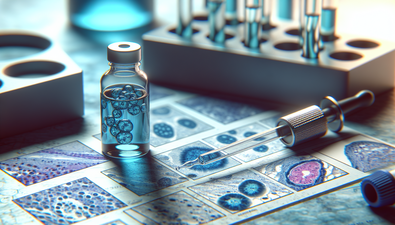
Methylene Blue is a synthetic dye that has been used in histology for over a century. Its chemical structure consists of a thiazine core, which is responsible for its strong staining capabilities. The dye is not only recognized for its ability to stain acidic cellular components, such as nucleic acids, but also for its utility in various histological applications.
The unique properties of Methylene Blue allow for detailed visualization of cellular structures, making it an invaluable tool in both basic and clinical research. It often serves as a counterstain in multi-step staining protocols, adding depth and contrast to histological slides.
Why Choose Methylene Blue for Histological Staining?
You might question the advantages of using Methylene Blue over other stains available. Its robustness and relative safety are two primary factors. Unlike some dyes that can be toxic or less reliable, Methylene Blue is known for its stability and ease of use. Additionally, it provides clear contrast in a wide range of biological specimens, from animal tissues to cellular cultures.
Notably, Methylene Blue is cost-effective and readily available, making it a practical choice for laboratories operating on a budget or in resource-limited settings. Its compatibility with various mounting techniques and imaging systems further increases its appeal.
Techniques for Optimal Staining with Methylene Blue
Let’s delve into specific techniques that enhance the effectiveness of Methylene Blue staining in histology. Mastery of these methods will ensure high-quality slides and reproducible results.
Standard Staining Protocol
The most fundamental approach to using Methylene Blue is the standard staining protocol. By adhering to a straightforward methodology, you can achieve reliable results.
-
Preparation of Tissue Samples: Ensure that your tissue samples are fixed properly, typically using formalin or another suitable fixative. After fixation, you should embed the samples in paraffin and prepare thin sections for mounting.
-
Deparaffinization: To prepare the slides, deparaffinize the tissue sections using xylene or a suitable substitute. Immerse the slides in decreasing concentrations of ethanol to rehydrate the sample.
-
Staining Process: Stain the section with Methylene Blue for approximately 5–10 minutes. You may wish to agitate gently to ensure even distribution of the dye.
-
Rinsing: After staining, rinse the sections in distilled water to remove excess dye.
-
Mounting: Mount the slides with appropriate media, such as a mounting medium that is compatible with microscopy.
This standard protocol is adaptable depending on the specific requirements of your tissue type and the details of your study.
Time and Temperature Considerations
Timing and temperature play critical roles in the effectiveness of Methylene Blue staining. Here are some points to consider:
-
Staining Duration: A staining duration of 5–15 minutes is typical, but optimization may be necessary based on the tissue type. More delicate samples may require shorter staining times to prevent excessive dye uptake.
-
Incubation Temperature: Performing the staining at room temperature is generally standard practice. However, slight warming may enhance the dye’s interaction with cellular components. Just be cautious not to exceed temperatures that could compromise the viability of the sample.
Adjusting these variables will allow you to tailor the protocol to specific research needs.
Enhancing Staining Intensity
If you find that your Methylene Blue staining lacks intensity, consider the following modifications:
-
Concentration Adjustments: Increasing the concentration of Methylene Blue can enhance staining. A 0.5% solution is standard, but higher concentrations may be beneficial for certain tissues.
-
pH Modifications: The staining efficacy can be improved by adjusting the pH of the staining solution. Methylene Blue is most effective at a slightly alkaline pH of around 7.0 to 9.0.
-
Incubation Time Extension: Lengthening the incubation time can amplify the staining intensity, but you must monitor the sections closely to avoid overstaining.
Experiment with these modifications to achieve the desired intensity for your histological slides.

Methylene Blue in Combination Stains
Methylene Blue is often utilized as a counterstain or in combination with other histological stains. Understanding how to effectively pair it with other reagents can yield insightful results.
Examples of Combination Stains
-
Methylene Blue and Eosin: This classic combination provides a comprehensive view of tissue structure. Eosin stains cytoplasmic components red, while Methylene Blue highlights the nuclei. This dual approach allows for better differentiation of cellular constituents.
-
Methylene Blue and Hematoxylin: Another effective pairing, particularly in the examination of cellular structures and nucleic acids. Hematoxylin provides a rich blue stain that complements Methylene Blue’s properties and enhances overall contrast.
-
Methylene Blue with Gram Stain: Employing Methylene Blue in Gram staining can help in differentiating Gram-positive and Gram-negative bacteria. The dye enhances the visibility of cellular morphology and arrangement.
Using Methylene Blue in combination can lead to more comprehensive assessments of the tissues under investigation.
Staining Protocols for Combination Stains
When implementing combination protocols, you must ensure that the sequences of staining do not compromise the quality of results.
-
Preparation: As with standard staining, begin with appropriately fixed and embedded tissue sections.
-
First Stain Application: Apply the primary stain (e.g., Hematoxylin) first. Follow the specific protocol for that stain regarding timing and rinsing.
-
Secondary Stain Application: After rinsing, apply Methylene Blue. Adjust the staining time based on results from previous trials for optimal differentiation.
-
Final Rinses and Mounting: As with other processes, rinse thoroughly and mount using a compatible mounting medium.
Troubleshooting Combination Stains
In some cases, staining combinations may not yield the desired outcome. Here are some troubleshooting tips:
-
Check Stain Compatibility: Ensure that the first stain does not interfere with the Methylene Blue staining process. Review literature on specific combinations for guidance.
-
Analyze Staining Times: Review the times used for each stain. A brief exposure to a stronger dye may wash out components needed for Methylene Blue visualization.
-
Consider Tissue Fixation: Inadequate fixation procedures can result in poor staining. Ensure that your fixation process is thorough and that tissue samples are adequately penetrated by the reagents.
Your careful attention to these details will improve your staining outcomes.
Applications of Methylene Blue in Histology
Methylene Blue’s versatility extends beyond mere aesthetic purposes in histology. It also serves crucial scientific roles in various applications.
Cellular Morphology Studies
Methylene Blue is frequently employed in studies of cellular morphology, helping researchers visualize structures within cells more clearly. Its affinity for nucleic acids allows for the easy identification of nucleus shape and size—factors that can indicate cellular health or pathology.
Viability Assays
In cell culture studies, Methylene Blue can serve as a reliable indicator of cell viability. Living cells uptake the dye, while dead cells do not, allowing researchers to assess cell health quantitatively.
Diagnostic Applications
Clinicians often utilize Methylene Blue for diagnostic procedures—such as identifying certain bacterial infections or evaluating tissue biopsies. Its clarity and detail enable pathologists to make accurate assessments of health conditions.
By understanding these applications, you can better appreciate the utility of Methylene Blue beyond traditional histological staining.

Best Practices for Working with Methylene Blue
When integrating Methylene Blue into your histological work, adhering to best practices can enhance outcomes and ensure safety.
Safety Measures
Always prioritize safety when working with chemicals. Methylene Blue, while relatively safe, can cause irritation upon contact with skin or eyes. Ensure that you monitor personal protective equipment and work in a well-ventilated area.
Optimal Storage Conditions
Methylene Blue should be stored in a cool, dark place to preserve its integrity. Exposure to light can degrade the dye, leading to inconsistencies in staining.
Keep Records
Documenting your staining protocols, observations, and results can provide insights into ongoing projects. Over time, these records can help optimize your practices and reinforce successful techniques.
Incorporating these best practices will help ensure the reliability and safety of your histological work with Methylene Blue.
Conclusion
The application of Methylene Blue in histological staining is a dynamic and rewarding practice that provides clear insights into cellular structures. By mastering the diverse techniques outlined in this article, you can enhance both the quality of your slides and your understanding of cellular pathology. Whether employed in standard protocols or combination techniques, Methylene Blue offers a reliable solution for detailed tissue analysis.
As you advance in your histological pursuits, remember that experimentation and adaptation are key to perfecting the art of staining. Implement these methods and cultivate a deeper appreciation for the intricacies of microscopy, armed with the knowledge to leverage Methylene Blue effectively.